
Which dental imaging equipment is the best choice for you?
In recent years, China’s dental industry has rapidly expanded, with the market size nearly doubling. As the sector matures, competition among dental institutions has intensified, with orthodontic and implant services now accounting for over 70% of the market share.
Both public hospitals and private clinics benefit from high patient volumes in these areas. To remain competitive, equipping dental practices with advanced imaging technology and digital solutions is essential.
We’ll be looking at all the essential equipment, from your standard X-rays right up to those fancy 3D scanners. Our goal is to help dental teams figure out the best tools for tackling all sorts of cases they see in the clinic.
Oral Radiology
Oral radiology, also known as “Oral and Maxillofacial Medical Imaging,” evolved from early dental radiology. Following the discovery of X-rays in 1895, they were quickly applied to dental films. Modern dental imaging devices typically include intraoral X-ray machines, panoramic X-ray machines, and dental-specific CT scans (CBCT). Additionally, oral scanners are used in orthodontics to create 3D models of the teeth, aiding in treatment planning.
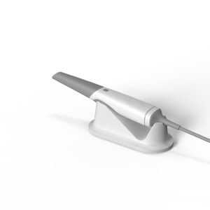
Oral Scanner
However, oral scanners do not include X-ray generating devices and can only produce images of the external surface of the teeth. They cannot penetrate the teeth to generate images of the teeth or the internal structures of the mouth. Therefore, oral scanners are typically not included in dental imaging equipment. Now, let’s break down how intraoral X-ray machines, panoramic X-ray machines, and CBCT (Cone Beam Computed Tomography) actually work – and, more importantly, how you can use them in your practice.
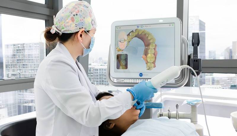
Intraoral X-ray machine
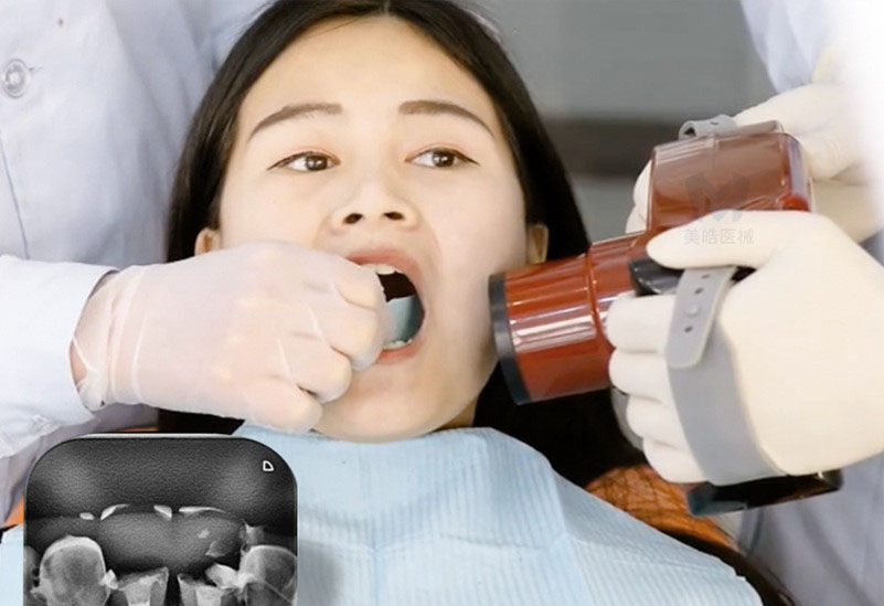
The doctor is using the intraoral X-ray machine to take a shot
An intraoral X-ray machine consists of an X-ray generating device and its supporting components. It has a simple principle and a compact size. During imaging, a film or detector is placed inside the mouth, behind the teeth, and the machine captures the image. Typically, it can capture 1 to 4 teeth at a time, providing a two-dimensional image of the interior of the teeth.
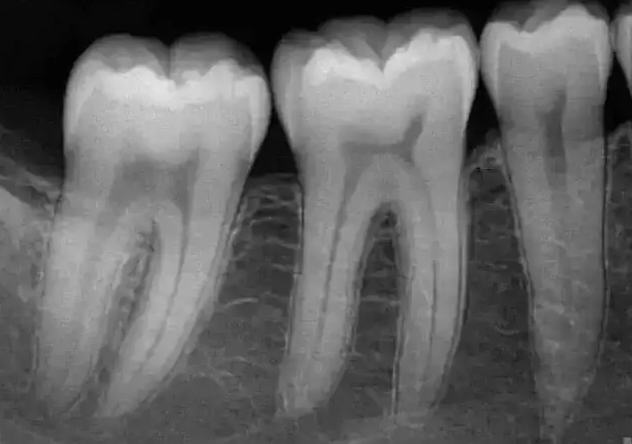
Intraoral X-ray machines are easy to use and provide quick results. They can clearly display both the crown and root of the teeth, making them commonly used for simple dental examinations such as detecting cavities, root fractures, root cysts, as well as assessing the effectiveness of dental fillings and root canal treatments.
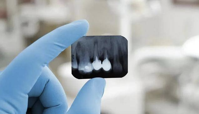
Small X-ray Film
As an affordable and basic dental imaging device commonly used in many dental institutions, intraoral X-ray machines do have their limitations. Due to their small imaging field, intraoral X-ray machines are nearly unable to capture images of the external surfaces of the teeth. As a result, they are generally not used for orthodontic or implant-related diagnostics and treatments.
Panoramic X-ray machine
A panoramic X-ray machine, also known as a “curved layer X-ray machine,” uses panoramic radiography technology to capture a comprehensive two-dimensional image of the entire mouth, including the teeth, periodontal tissues, and adjacent anatomical structures, with a single exposure. The resulting image covers the entire oral cavity, much like a “group photo” of the teeth.
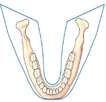
Panoramic X-ray Imaging Range
Think of panoramic X-rays as the dentist’s go-to for getting the ‘big picture’ view of your mouth. They’re incredibly useful for picking up on signs of significant tooth loss, potential issues within the jawbone itself (like cysts or tumors), fractures from injuries, or even developmental problems that might affect the growth and alignment of teeth. And, for those considering braces or other orthodontic work, panoramic X-rays give dentists essential information to create the best treatment plan.

Panoramic X-ray Image
Compared to intraoral X-rays, the clarity and detail of the internal structures of the teeth in panoramic images are somewhat inferior. Additionally, because panoramic X-ray imaging uses curved layer technology, the resulting images often suffer from significant distortion and may produce overlapping ghost images. As a result, in dental services such as implantology, where high clarity of tooth structure imaging is crucial, it is difficult to rely solely on panoramic X-rays to formulate accurate treatment plans.
Dental CBCT (Cone Beam Computed Tomography)
In the previous article of this series, we introduced the imaging principles of CBCT . In simple terms, dental CBCT uses a large-area flat panel detector to receive cone-shaped X-ray beams emitted from an X-ray source, thereby generating accurate and clear three-dimensional images of the oral and maxillofacial structures.
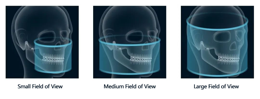
Today’s CBCT technology can capture three types of imaging fields: small field of view, medium field of view, and large field of view.
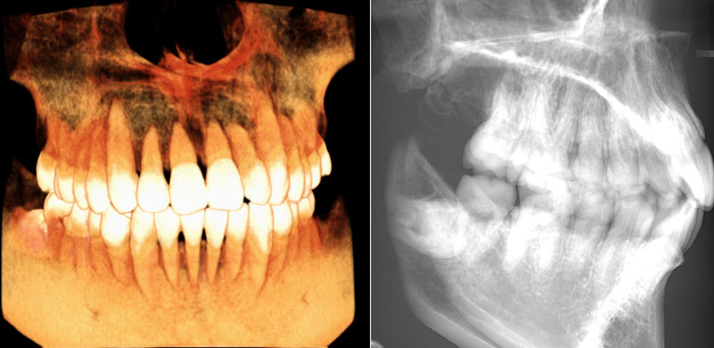
Small Field of View Image
A small field of view can capture a portion of the upper and lower teeth, along with the dental arch. However, it cannot display both the bilateral temporomandibular joints or the third molars on both sides in a single image. Dentists commonly use it for detailed diagnostics such as root canal treatments, root fractures, and dental microfractures.
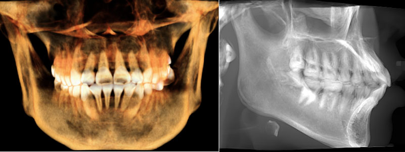
Medium Field of View Image
A medium field of view can fully display the entire dental arch. However, it cannot cover the full image of both temporomandibular joints or the entire maxillary sinus. It is more suitable for clinical applications such as single or multiple dental implants. It is also useful for observing and diagnosing the maxillary sinus floor, bilateral temporomandibular joints, and parts of the maxillary sinus contours.
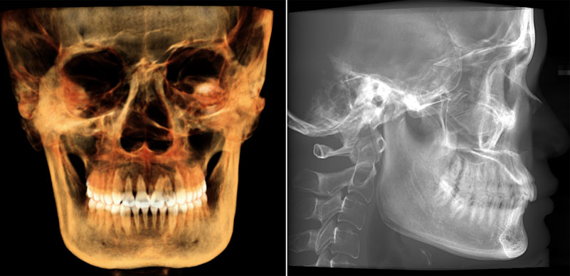
Large Field of View Image
A large field of view can cover most of the craniofacial region. It shows the complete morphology from the upper to the lower jaw, including both temporomandibular joints. This makes it ideal for several clinical applications. These include full dental arch imaging, maxillofacial surgery, full-mouth implants, and orthodontics and orthognathic surgery.
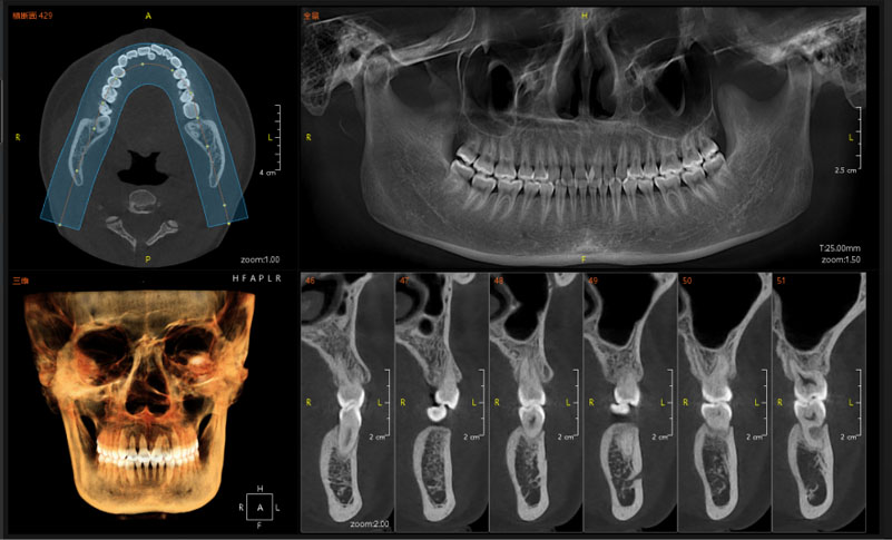
CBCT Imaging Software
One of the major advantages of CBCT is that it typically includes specialized dental imaging software with the equipment. The software’s built-in features can automatically generate panoramic images without overlapping ghost images. Additionally, it allows for image segmentation to obtain “small X-rays” of individual or multiple teeth. The software automatically corrects the processed images for distortion, providing clearer images with more detailed information. Moreover, the software allows rotation and observation within a three-dimensional plane. This makes it an excellent alternative to both intraoral X-ray machines and panoramic X-ray machines.
CBCT offers significant advantages in terms of image quality, operational modes, patient experience, and clinical applications. Regarding the commonly concerned issue of radiation dose, the radiation levels of CBCT are also within an absolutely safe range.