
Occlusal Surface Scanning
Initiate the scan by positioning the scanner parallel to the occlusal surfaces of the molars. Maintain steady, overlapping movements to capture detailed fissures and cusps. As you transition to the anterior region, tilt the scanner slightly toward the lingual aspect to avoid obstructing the scanning path. For those learning how to use intraoral scanner effectively, ensure the device consistently faces the patient’s throat, with the entire arch centered in the viewfinder. Smooth, continuous motion is critical—hesitation may cause stitching errors.
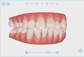
Lingual Surface Scanning
Approach buccal surfaces in two segments: molars to midline on both sides. Angle the scanner at ~45° relative to the occlusal plane for posterior teeth. How to use intraoral scanner in tight anterior spaces: retract the cheek with your non-dominant hand and hold the scanner horizontally, angling the tip toward the scanning direction. This minimizes shadowing near cervical margins.
Buccal Surface Scanning
Approach the buccal surfaces in two segments: from the molars to the midline on both sides. For posterior teeth, angle the scanner at approximately 45° relative to the occlusal plane, ensuring simultaneous visualization of buccal grooves and occlusal edges. In the anterior region, position the scanner closer to the tooth surface while retracting the cheek with your non-dominant hand. Adjust your grip: hold the scanner horizontally, angling the tip toward the scanning direction (e.g., tip left for left quadrants). This minimizes shadowing and optimizes access to cervical margins.

Incisal Edge Capture
For anterior teeth, pivot the scanner from the lingual to the buccal aspect in a “flipping” motion to trace the entire incisal edge. Keep the edge centered in the viewfinder while using a finger to retract the lip. If incisal wear or chips are present, perform a supplemental slow-motion pass to enhance detail. Adjust lighting if the scanner permits—reducing glare on enamel improves edge detection.
Interproximal and Bite Registration
To record occlusion, position the scanner midway between the arches. Capture posterior quadrants first using a wave-like motion, moving from molars to canines while maintaining contact with the buccal surfaces. Instruct the patient to lightly close their teeth but avoid forceful biting. For stubborn stitching gaps, rescan overlapping zones with slower, shorter strokes.
Distal Surfaces of Terminal Molars
Extend the scanner posteriorly beyond the last molar, tilting the handle downward to visualize the distal surface. Rotate the head alternately toward the buccal and lingual aspects to composite a full 3D model. If tissue obstruction occurs, use a mirror or cotton roll to depress the retromolar pad. Verify distal contours in real-time—incomplete data here frequently necessitates retakes.
Data Validation Protocol
Before finalizing, systematically inspect the virtual model:
Anterior Integrity: Confirm incisal edges, lingual concavities, and labial surfaces are seamless. Check for “ghosted” areas caused by lip movement.
Posterior Coverage: Ensure all occlusal pits, buccal grooves, and disto-lingual cusps are fully rendered. Zoom in to verify interproximal contacts lack voids.
Gingival Margin Accuracy: A minimum 2mm gingival cuff must surround each tooth. Blurred or truncated soft tissue suggests repositioning errors.
Bite Alignment: Use the software’s articulation tool to confirm intercuspation matches clinical observations. Mismatched fossa-to-cusp relationships indicate scanning sequence errors.