Common Types of Dental X-Ray Machines
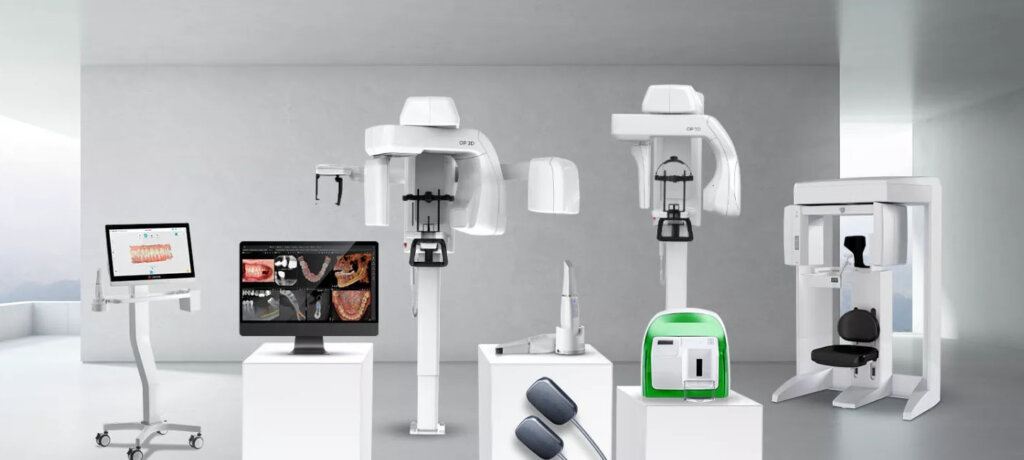
Dental X-ray machines can be categorized into the following main types:
- Traditional X-Ray Machines
These are the most common type, using film to capture images. While they provide high-quality imaging, their operation can be more cumbersome and requires additional time for processing. - Digital X-Ray Machines
Compared to traditional systems, digital X-ray machines offer clearer and faster imaging. They allow images to be processed and stored digitally, significantly improving efficiency and ease of use. - Panoramic X-Ray Machines
This type of equipment captures the entire oral cavity in a single image. It enables dentists to get a comprehensive view of the patient’s oral health, making it particularly useful for initial assessments. - CBCT (Cone Beam Computed Tomography)
CBCT uses advanced imaging technology to provide 3D images, allowing for more precise diagnostics. This type of machine is especially valuable for complex cases requiring detailed anatomical analysis.
Basic Components of a Dental X-Ray Machine
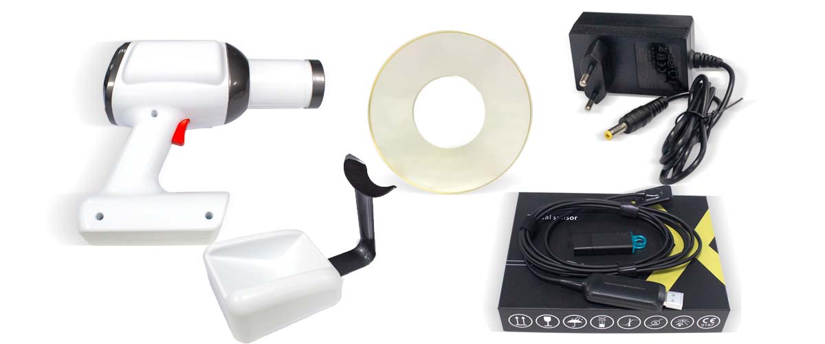
Before diving into the operation process, it’s essential to understand the primary components of a dental X-ray machine. Typically, the machine consists of the following key parts:
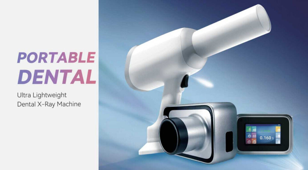
- X-Ray Emitter: Produces X-rays to capture images of the oral cavity.
- Receptor: Receives X-rays that pass through oral tissues and converts them into visible images.
- Control Panel: Used to set parameters and functions for imaging.
- Stand and Base: Supports the device and ensures stability during operation.
Operating a Dental X-Ray Machine
1. Preparation
- Check the Equipment: Before starting, ensure the X-ray machine is functioning properly and has no defects.
- Prepare Protective Gear: Provide lead aprons and thyroid collars as needed to protect patients from radiation exposure.
2. Communicate with the Patient
- Obtain Medical History: Verify if the patient has allergies, pregnancy, or other conditions to ensure safety.
- Explain the Procedure: Describe the purpose and steps of the X-ray process to ease patient anxiety.
3. Position the Patient
- Adjust the Chair: Ensure the patient is seated correctly for proper imaging.
- Guide the Patient: Use tools like bite blocks to help the patient maintain the correct position.
4. Set Up the Equipment
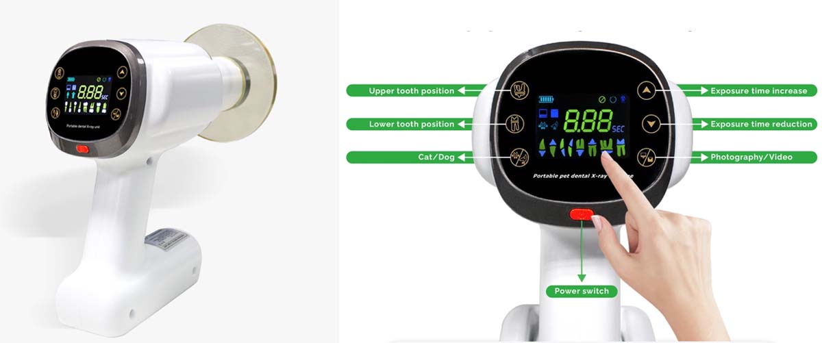
- Select Imaging Type: Choose the appropriate imaging mode, such as panoramic, bitewing, or periapical.
- Adjust Parameters: Set the exposure time and current on the control panel to achieve high-quality images.
5. Perform the Imaging
- Ensure Safety: Confirm all protective measures are in place before taking the X-ray.
- Capture the Image: Press the imaging button to emit X-rays, minimizing radiation exposure time.
6. Review the Images
- Inspect Image Quality: Immediately check the images to ensure they clearly display the required details.
- Retake if Necessary: If the images are unclear, conduct a second imaging session as needed.
7. Organize and Record
- Save Images: Store the images in the patient’s electronic health records for future reference.
- Document Details: Record the imaging date, type, and relevant notes for subsequent use.
Tips to Enhance Efficiency
Once familiar with the basic operation process, the following tips can help dental professionals improve their efficiency when using X-ray machines:
- Training and Practice
- Conduct regular training sessions for dentists and assistants to ensure everyone is proficient in operating the equipment.
- Streamline Workflow
- Organize tasks to ensure each step transitions smoothly, minimizing unnecessary delays.
- Leverage Digital Imaging
- Use digital X-ray systems to quickly obtain images and process them on computers, enhancing overall efficiency.
- Regular Equipment Maintenance
- Schedule periodic maintenance and calibration of the X-ray machine to keep it in optimal condition and avoid disruptions caused by malfunctions.
- Establish Standardized Procedures
- Develop and follow standardized workflows to reduce human error and improve overall operational efficiency.
By mastering the operation process and adopting these practical tips, dental professionals can maximize the utility of X-ray machines, ensuring both efficiency and high-quality patient care.


