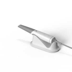

How Does An Intraoral Scanner Work: A Deep Dive into the Technology Revolutionizing Modern Dentistry
Intraoral scanners have transformed dental practices by replacing traditional, messy impression materials with precise digital workflows. But how does an intraoral scanner work to capture intricate details of teeth and gums in seconds? Let’s unpack the engineering magic behind these tools, layer by layer.
1. Triangulation Technology: The Backbone of Precision
Triangulation isn’t just for land surveying—it’s the unsung hero of intraoral scanning. Here’s how it works:
- A narrow beam of light (often blue or green LED) is projected onto the tooth surface.
- As the light hits uneven surfaces—like a molar’s fissures or a crown’s margin—it scatters. A miniature camera (typically a charge-coupled device (CCD) or CMOS sensor) captures this distorted light pattern.
- Using geometric trigonometry, the scanner calculates the distance between the light source, the reflected point, and the sensor. Even a 0.01mm groove creates measurable shifts in the light’s path.
Why it matters: This method achieves micron-level accuracy—critical for detecting early caries or ensuring crown margins fit like puzzle pieces. Early models like the CEREC Omnicam relied heavily on this approach, though modern systems combine it with other tech for better results.

2. Parallel Confocal Imaging: Cutting Through the Clutter
Ever tried taking a photo in fog? Traditional optics struggle with saliva or blood, but confocal imaging laughs in the face of moisture. Here’s the breakdown:
- The scanner emits parallel laser beams at specific wavelengths (often red or near-infrared).
- Only light reflected from the exact focal plane passes through a microscopic pinhole filter. Everything else—blurry reflections from saliva, cheek tissue, or blood—gets blocked.
- By rapidly adjusting the focal distance (up to 300 times per second!), the device builds a “depth map” of the entire arch.
Real-world impact: This is why scanners like the iTero Element can work even if a patient moves slightly mid-scan. It’s like having an autofocus system that’s 100x faster than your smartphone camera.
3. Laser Triangulation: Speed Meets Detail
Think of this as triangulation 2.0. Instead of static light patterns, here’s where things get dynamic:
- A red laser line sweeps across teeth at 20,000 oscillations per second, guided by a vibrating micro-mirror.
- Multiple cameras (usually 3-5) surround the scanner tip, capturing the laser’s distortion from different angles.
- Advanced algorithms stitch these perspectives into a cohesive 3D model.
Industry example: The 3Shape TRIOS 4 uses this method to capture a full arch in under 60 seconds. Dentists love it for orthodontic cases—imagine tracking 0.2mm tooth movements across six months of Invisalign treatment!
4. Structured Light + Laser Fusion: The Best of Both Worlds
Why choose one technology when you can hybridize? Newer scanners like the Medit i700 blend structured light with laser triangulation:
- Structured light: Projects a grid of microscopic dots (think 50,000 dots per frame). Distortions in this grid reveal surface contours.
- Laser backup: When blood or deep shadows confuse the structured light system (e.g., in a subgingival prep), lasers provide fallback data.
Clinical advantage: This combo conquers “problem zones” like second molars or edentulous ridges. A 2023 study in J Prosthodontics showed hybrid systems reduce rescan rates by 63% compared to single-tech devices.
Beyond the Scanner: How Data Becomes a Crown
Capturing the scan is just step one. Here’s the behind-the-scenes workflow most patients never see:
- Mesh generation: Raw data points (often 10+ million per scan) get converted into a 3D polygon mesh.
- AI cleanup: Algorithms remove “noise” like transient bubbles or tongue artifacts.
- Alignment: Upper and lower arches are digitally articulated using bite registration scans.
- Design: CAD software (ex: exocad) lets technicians sculpt restorations that align with the patient’s occlusion.
- Fun fact: Some systems now integrate 3D printing simulations, showing how layer-by-layer fabrication will occur—a godsend for explaining treatment plans to anxious patients.
Why Dentists Are Ditching Impressions

- Accuracy: Studies show intraoral scanners achieve 15-30μm precision vs. 60-100μm for PVS impressions.
- Comfort: No more gag-inducing trays—a 2022 survey found 89% of patients prefer scans.
- Efficiency: Same-day crowns are now routine. A scan-to-mill workflow can deliver a ceramic restoration in 15 minutes.
But it’s not all roses. Challenges persist:
Subgingival margins: Scanning a prep 1mm below the gumline still requires retraction cords or AI guesswork.
Cost: High-end scanners like the Primescan run ~$25k—though leasing options are easing the pain.