
How Dental X-Ray Machine Works: A Comprehensive Guide
What is an X-ray?
X-rays are a form of electromagnetic radiation, similar to visible light but with much higher energy.
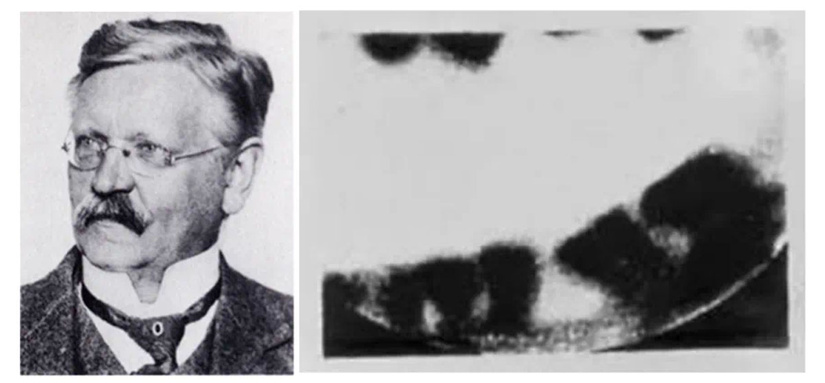
Dental radiology is the full name of “oral and maxillofacial medical imaging”, which has gradually evolved from the original “simple dental radiology”. After X-rays were first discovered by German physicist Wilhelm Conrad Roentgen in 1895, they were soon used in dental X-rays. In early 1896, German dentist Otto Walkhoff exposed the first intraoral radiographic image.
Unlike visible light, which most objects absorb or reflect, X-rays pass through soft tissues while denser materials like bones and teeth absorb them. They allow healthcare professionals to see inside the body without invasive procedures.
How Does a Dental X-Ray Machine Work?
A dental X-ray machine operates by emitting controlled bursts of X-ray radiation to capture detailed images of the teeth, bones, and surrounding oral structures. The process involves several key components and steps:
- X-ray Tube: This is the core of the machine where electrons are generated and accelerated towards a metal target (usually tungsten). When the high-speed electrons collide with the target, X-rays are produced.
- Collimator and Filter: The collimator focuses the X-ray beam to direct it precisely at the area of interest, reducing exposure to surrounding tissues. A filter removes lower-energy X-rays that do not contribute to imaging quality.
- Receptor (Film or Digital Sensor): Traditional X-ray machines use photographic film, while modern ones utilize digital sensors to capture images. Digital sensors provide faster results, lower radiation exposure, and enhanced image quality.
- Image Processing: Once captured, the system either develops the image (in traditional film X-rays) or digitally processes it using specialized software. Digital images can be enhanced, magnified, and shared electronically for better diagnosis and record-keeping.

how dental x ray machine works
Why Do We Need Dental X-Ray Machines?
Dental X-rays play a crucial role in oral healthcare by allowing dentists to diagnose conditions that are not visible during a standard oral examination. Some key benefits include:
- Early Detection of Cavities: X-rays help identify tooth decay between teeth or beneath existing fillings that might otherwise go unnoticed.
- Assessment of Tooth and Bone Health: X-rays reveal issues like bone loss due to gum disease, impacted teeth, and root infections.
- Treatment Planning: Whether for orthodontics, dental implants, or root canals, X-rays provide vital information for creating personalized treatment plans.
- Monitoring Growth and Development: Pediatric dentists use X-rays to track the development of children’s teeth, identifying potential problems early.
Without dental X-ray machines, many oral health issues would remain undiagnosed until they cause significant pain or damage, leading to more complex and costly treatments.
How Do Dentists Ensure Your Child’s X-Ray Exam Is as Safe As Possible?
Radiation exposure is a common concern for parents when their children need dental X-rays. However, advancements in technology and strict safety protocols have made dental radiography safer than ever. Here’s how dentists minimize risks:
- Use of Low-Radiation Digital X-Rays: Modern digital sensors require significantly lower radiation doses compared to traditional film X-rays.
- Lead Aprons and Thyroid Collars: Protective gear shields sensitive body parts from unnecessary exposure.
- Selective Imaging: Dentists only take X-rays when absolutely necessary, based on a patient’s oral health history and risk factors.
- High-Speed Film or Digital Sensors: Faster image capture reduces exposure time, minimizing radiation absorption.
- Proper Technique and Positioning: Ensuring accurate X-ray positioning prevents retakes, further limiting exposure.
When parents bring their kids in for checkups, dental teams follow strict safety playbooks developed by the ADA and medical oversight boards. Those child-specific protocols – like adjustable radiation doses and lead apron placements – are why generations have grown up with X-rays as routine as fluoride treatments.
This tech revolution didn’t happen by accident. Equipment manufacturers work hand-in-hand with dental associations, rolling out smarter digital systems that help clinicians map out root canals like GPS navigation while cutting exposure levels annually. Dentists now complete in single shots what once required multiple retakes, reducing chair time for fidgety patients. The result? Safer scans and more accurate treatment plans that keep our pearly whites actually white.