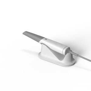

Why Microscope Positioning Starts with Perfecting Dental Chair Positions
With domestic dental microscopes now rivaling global brands in precision and affordability, clinics are rapidly adopting them for root canals, aesthetic procedures, and apicoectomies. But here’s the catch many overlook: dental chair positions directly determine whether your $20,000 microscope becomes a game-changer or a paperweight.
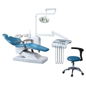
Think about it—during a molar root canal, if the patient’s head tilts just 10° off the ideal dental chair position, your microscope’s focal depth gets compromised, forcing constant refocusing that adds minutes per procedure. This guide cuts through the frustration by linking three dental chair positions (supine, semi-reclined, upright) to specific microscope angulations.
For example:
- Supine dental chair position: Raise the microscope 20° from horizontal to visualize palatal canals without neck strain.
- Aesthetic cases: Combine 45° chair recline with lateral microscope tilt to capture incisal edge translucency.
We’ve mapped these setups using real-world data from 37 clinics—because mastering microscopy isn’t about the gear alone, but how you synchronize it with dental chair positions that unlock its full potential.
Operating position of dental microscope
When using the microscope, the objective lens should generally be kept basically vertical to the ground (as shown below). The mouth mirror is at a 45° angle to the direct light path, and the binoculars are at an angle of 165°~185° to the ground. The best viewing angle is achieved through the reflection of the mouth mirror. When treating maxillary teeth, the plane of the mouth mirror is at a 45° angle to the ground; when treating mandibular teeth, the plane of the mouth mirror is at a 120° angle to the ground. Depending on the treatment content and tooth position, the operating position of the microscope can be adjusted accordingly. Look at the proper position and mirror angle of the maxillary dental arch
Look at the proper position and mirror angle of the mandibular dental arch
Working position of the maxillary anterior area
1. Doctor: Model at 12 o’clock;
2. Assistant: 2-4 o’clock, hold the strong suction tube in the left hand and insert it into the patient’s mouth at the right corner of the mouth;
3. Microscope: The mirror body is perpendicular to the ground;
4. Patient: Supine, the maxillary plane is perpendicular to the ground or at a 45° angle.

Working position of the upper left posterior area
1. Dental chair: Slightly raise the dental chair and adjust it so that the patient’s mandibular plane is parallel to the ground plane and the maxillary plane is at a 45° angle to the ground plane;
2. Doctor: 11-12 o’clock;
3. Nurse: 3 o’clock, put the strong suction tube tip into the patient’s mouth;
4. Microscope: Reduce the angle between the microscope and the axial plane where the root canal is located;
5. Patient: Supine, turn the head slightly to the right when treating premolars, and turn the head completely to the right when treating molars. Slightly toward the left side.

Working position in the right upper posterior teeth area
1. Dental chair: slightly raised, adjusted to make the patient’s mandibular plane parallel to the ground plane, the maxillary plane at a 45° angle to the ground plane, and the surgical area under the operating microscope;
2. Doctor: 11 o’clock position, 65° angle with the patient or the long axis of the dental chair;
3. Assistant: 2 o’clock to 3 o’clock position, the strong suction tube is inserted into the patient’s mouth at the left corner of the mouth, the head of the suction tube is placed on the back of the lower posterior teeth, and the left little finger is fixed on the patient’s chin;
4. Microscope: reduce the angle between the microscope and the axial plane where the root canal is located;
5. Patient: supine, head tilted back; when treating the right upper premolar, the patient’s head should be slightly toward the left; when treating the right upper molar, the patient’s head is completely toward the left to facilitate the doctor to operate in the right upper maxillary posterior teeth area, and the chin is extended to the right side.

Working position in the lower left posterior area
1. Dental chair: adjust the chair position to a 30° angle between the backrest and the ground, and place the surgical area under the operating microscope;
2. Doctor: 10 o’clock to 12 o’clock position;
3. Assistant: 2 o’clock to 3 o’clock position, put the strong pipette tip into the patient’s mouth, and gently press the patient’s tongue to the lower right;
4. Microscope: increase the angle between the microscope and the axial plane where the root canal is located; 5. Patient: lie on his back, slightly facing the right side.

Working position in the lower right posterior area
1. Dental chair: slightly raise the dental chair and adjust it to make the patient’s mandibular plane parallel to the ground plane, the patient’s mouth and elbow at the same level, and the backrest at a 40° angle to the ground plane;
2. Doctor: 7 o’clock to 9 o’clock position;
3. Assistant: 3 o’clock to 4 o’clock position, hold the patient’s chin with the ring finger, and gently press the patient’s tongue down with the strong pipette tip;
4. Microscope: increase the angle between the microscope and the axial plane where the root canal is located;
5. Patient: lie on his back, slightly facing the left side.